Nuclear Medicine & Molecular Imaging

Ruby Hall Clinic describes its Nuclear Medicine, Molecular Imaging and Theranostics Department as a long-standing advanced service established in 1996, and specifically notes that it was the first hospital-owned nuclear medicine department in Maharashtra outside Mumbai.
The mirrored source positions the unit as both a diagnostic and therapeutic specialty, spanning renogram studies, bone scans, PET/CT imaging, radionuclide therapy, theranostics, and specialised treatment pathways for thyroid disease, neuroendocrine tumours, prostate cancer, liver tumours, and chronic joint conditions.
A major differentiator on the source page is the in-house GE MINItrace Cyclotron, which supports on-site radiopharmaceutical production and helps the department deliver faster PET imaging access, targeted therapies, and more responsive personalised care.
Advanced nuclear medicine with molecular imaging, PET/CT precision, theranostics, and on-site cyclotron support for faster diagnosis and targeted treatment.
A Department Built Around Molecular-Level Diagnosis And Targeted Therapy
The department is presented as a specialty that goes beyond structural imaging by looking at physiological and molecular activity inside the body. This makes it especially valuable for early disease detection, treatment planning, response assessment, and long-term surveillance.
Ruby Hall Clinic also frames the service as a combined diagnostics-and-therapy platform, where imaging pathways such as FDG, DOTA, PSMA, FAPI, and Exendin PET scans sit alongside therapeutic programs such as radioiodine therapy, PRRT, TARE, radio ablations, and radiation synovectomy.
Why Choose Ruby Hall Clinic For Nuclear Medicine


Historic leadership in nuclear medicine
The mirrored source states that the department was established in 1996 and was the first hospital-owned nuclear medicine department in Maharashtra outside Mumbai.

Diagnostics and treatment in one specialty pathway
The department combines functional imaging, PET/CT, radionuclide therapies, theranostics, and preparation protocols inside one integrated clinical service.

On-site cyclotron advantage
In-house radiotracer production improves speed, supports time-sensitive PET workflows, and expands access to advanced imaging and therapy options.

Advanced tumour-specific imaging and therapy
The source highlights specialised pathways including DOTA, PSMA, FAPI, Exendin, PRRT, TARE, and radioiodine therapy for carefully selected patients.
Our Mission
- To deliver precise molecular-level diagnosis and targeted radionuclide therapy through an integrated nuclear medicine platform that improves treatment selection, response monitoring, and access to advanced personalised care.
Nuclear Medicine And Molecular Imaging Services

Diagnostic Nuclear Medicine
Functional scans such as renal studies, bone scans, and thyroid imaging help assess organ performance, tracer uptake, obstruction, and disease activity in ways conventional imaging cannot.
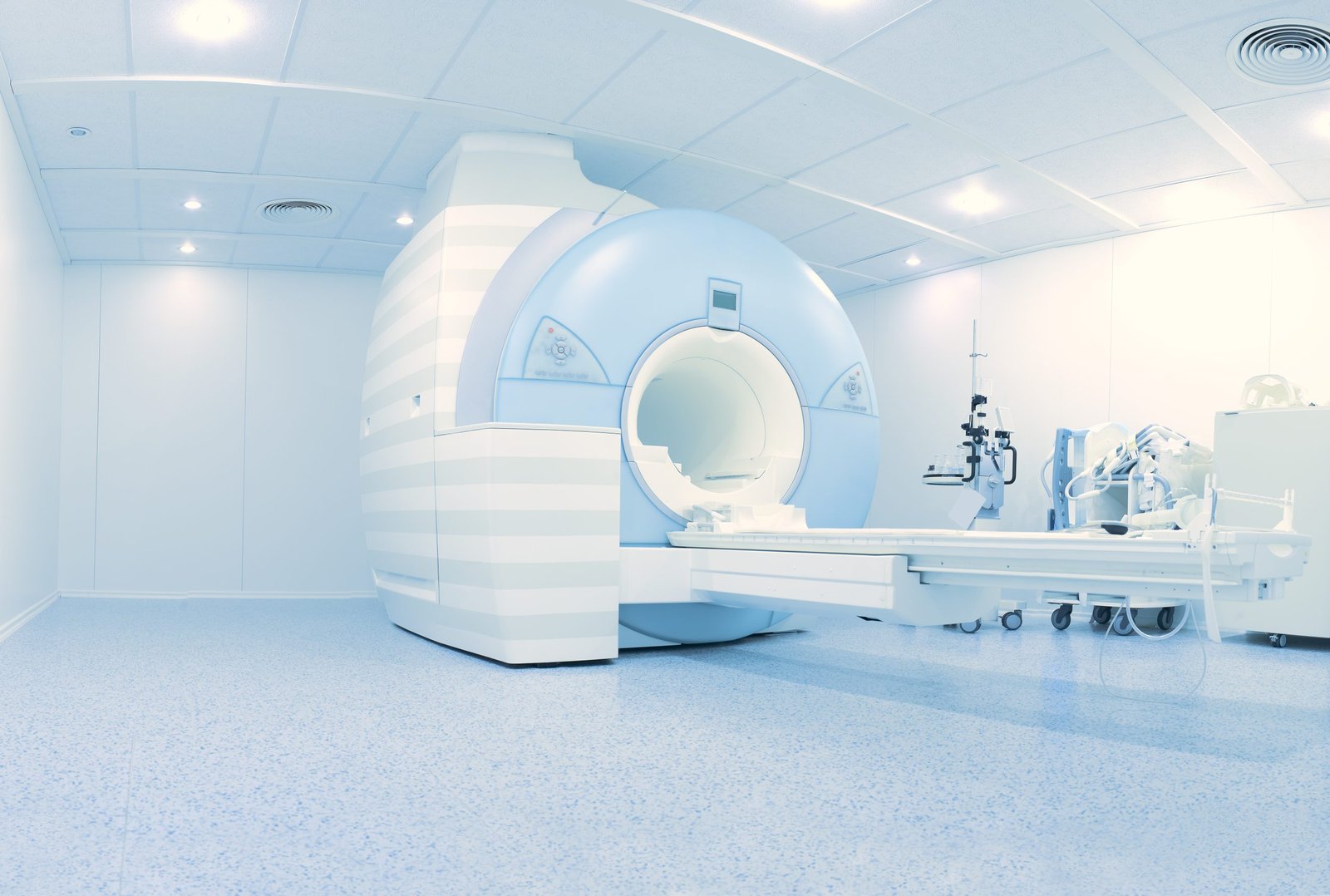
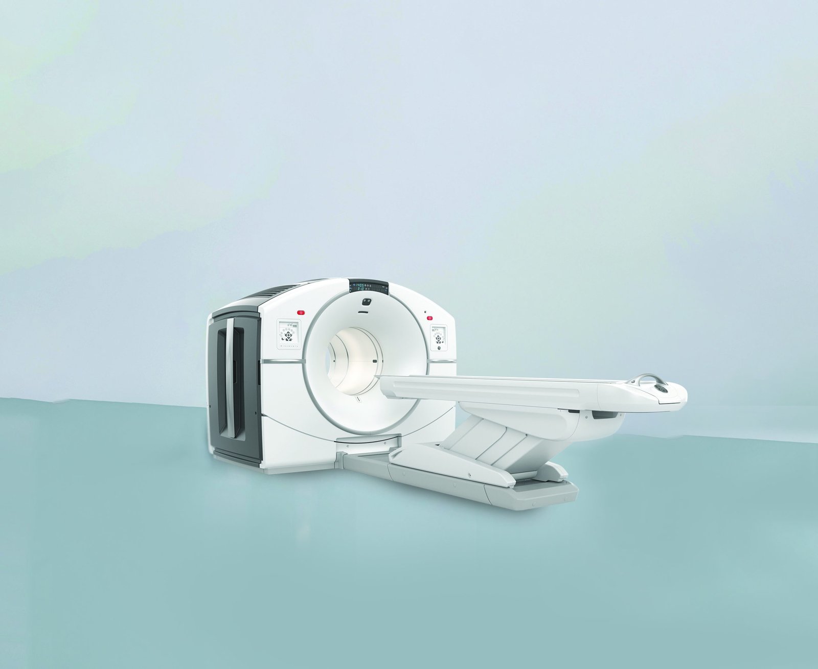
Molecular Imaging PET/CT
PET/CT programs include FDG, DOTA, PSMA, FAPI, and Exendin imaging for cancer detection, staging, recurrence assessment, treatment planning, and selected endocrine tumour localisation.
Theranostics
The department combines imaging and targeted treatment using tumour-specific radioisotopes for thyroid disease, neuroendocrine tumours, prostate cancer, liver tumours, bone pain palliation, and selected complex oncologic cases.

PRRT And Targeted Radionuclide Therapy
DOTA-PRRT, PSMA-directed therapy, and evolving FAPI-based approaches are used to treat receptor-positive tumours with internal radiation delivered directly to disease sites.
Cyclotron-Supported Radiopharmaceutical Access
The in-house GE MINItrace Cyclotron improves tracer availability, shortens delays, supports research and personalised medicine, and strengthens the turnaround for time-sensitive PET/CT studies.
Patient Preparation And Therapy Monitoring
The source emphasises preparation guidance, hydration, medication adjustment, blood and kidney-function checks, and post-treatment monitoring as core parts of safe nuclear medicine care.
Core Nuclear Medicine Programs
Ruby Hall Clinic uses diagnostic nuclear medicine to evaluate organ function and disease activity through tracer-based imaging rather than anatomy alone. The mirrored source highlights renal scintigraphy with DTPA or MAG3 to assess kidney function, drainage, obstruction, and transplant monitoring, along with additional diagnostic work in bone, thyroid, and other functional studies. The same service block also introduces radio ablations, TARE, radiation synovectomy, and early therapy workups, showing how the department blends diagnosis with intervention planning.
The molecular imaging section focuses on PET/CT as a way to see metabolic and cellular activity with anatomical correlation. FDG PET supports broad cancer, infection, brain, and cardiac evaluation; DOTA PET helps diagnose and stage neuroendocrine tumours and determine suitability for DOTA-PRRT; PSMA PET improves prostate cancer detection, staging, recurrence localisation, and treatment planning; FAPI PET is presented as an emerging option when FDG is less informative; and Exendin PET is used for insulinoma localisation and preoperative planning.

The theranostics program combines diagnosis and treatment using tumour-targeting radioisotopes. The source lists oral radioiodine for thyroid cancer and hyperthyroidism, PRRT for receptor-positive tumours, targeted IV therapies for neuroendocrine, prostate, and medullary cancers, liver-directed radioisotope treatment, bone pain palliation, and intra-articular therapy for synovitis and haemophilia-related joint disease. Patients may need pre-scan confirmation, hydration, medication adjustment, fasting, and short in-hospital isolation depending on the treatment path.
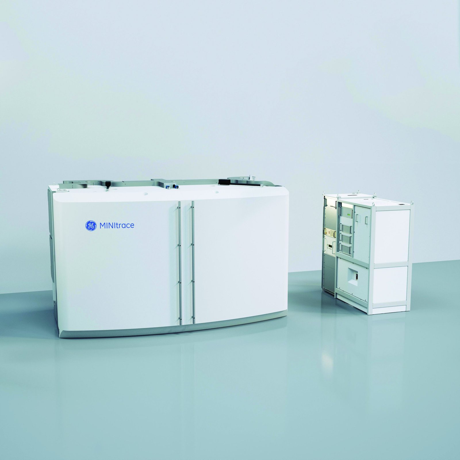
Ruby Hall Clinic highlights the GE MINItrace Cyclotron as Pune’s first such facility for on-site radioisotope production. It supports faster PET/CT access, improved tracer freshness, better imaging precision, and broader availability of specialised isotopes such as 18F-FDG, Gallium tracers for PSMA and DOTA imaging, 13N for selected advanced studies, and FAPI tracers for research-oriented and specialised applications. The page frames this capability as central to faster turnaround, personalised medicine, theranostics, and research support.
Nuclear Medicine FAQs
Is nuclear medicine safe?
Yes. The source states that radiation doses are generally low and carefully controlled, and that the benefit of accurate diagnosis or effective therapy outweighs the associated risk in properly selected patients.
What is the difference between a PET scan and a CT or MRI scan?
Ruby Hall Clinic explains that PET shows function and metabolic activity, while CT and MRI mainly show structure. PET/CT combines both to provide a more complete clinical picture.
Will I be radioactive after a scan or therapy?
For diagnostic studies, the residual radioactivity generally wears off within a few hours. After therapies such as PRRT or I-131, some patients may need isolation for one to two days depending on the treatment dose.
Can children or elderly patients undergo nuclear medicine procedures?
Yes. The department notes that nuclear medicine is used across age groups, with dose adjustments and protocol changes made to preserve safety.
How do I prepare for scans and therapies?
Preparation depends on the study. FDG PET usually requires 4 to 6 hours of fasting and blood sugar control, while many renal, bone, or thyroid scans need little or no fasting. Therapies often require blood and kidney-function testing, confirmatory imaging, medication adjustment, hydration, and in some cases temporary inpatient isolation.
How do I make an appointment?
The mirrored page directs patients to schedule through the department by calling 02066455265 or 02066455245 for Monday-to-Saturday appointments at the Super Specialty Building on the Ruby Hall Clinic campus.
Need PET/CT, Theranostics, Or Nuclear Medicine Advice?
An early specialist consultation can help determine whether a functional scan, tumour-specific PET study, or radionuclide therapy pathway is the right next step for diagnosis, staging, or treatment response planning.
Book an AppointmentPatient Preparation And Academic Depth
The page also stands out for two supporting strengths that many thinner department pages lack. First, it gives practical preparation guidance for both scans and therapies, including fasting rules, hydration, medication changes, confirmatory PET imaging, kidney-function testing, and pregnancy or breastfeeding precautions.
Second, the department is presented as an academic unit as well, with a DNB Nuclear Medicine programme offering primary and secondary seats each year. That combination of clinical depth, procedural guidance, and training capability makes the service feel more complete and institutionally mature than a standard imaging page.
Book an AppointmentMeet Our Doctors
Dr. Sameer Sonar
Nuclear Medicine and Molecular Imaging
- Mon to Sat (by appt)
- NA
Dr. Amol Gangadhar Galge
Nuclear Medicine and Molecular Imaging
- Mon to Sat
- 9:00am to 5:00pm (by appt)